All the microphotos seen on this website were photographed using various microscopes (seen in the photos below), some with integrated film cameras and also using a Nikon D-70 digital SLR with an eyepiece adapter. Almost all microphotos were taken at 20x - 100x magnification. Because microscopes operate at fixed focal lengths and have very shallow depth of field, focus is a difficult issue for microphotographs. When integrated cameras lacked film plane viewfinders, focus was accomplished using a proxy monocular installed in the t-mount or the binocular eyepieces. For film images, I used Fuji Provia 100 slide film and then scanned the slides at 3,200 dpi using an Epson 1680 scanner. Digital images were taken using the Nikon Camera RAW format (NEF) with auto white balance, and all images were processed in Adobe 1998 RGB color profile using Photoshop.
The dramatic colors associated with microphotos of rock thin sections and chemical crystals were obtained using a polarizing microscope, a standard tool used by petrographers, geologists who identify the mineralogy and textural properties of rocks. This technique uses plane-polarized light that interacts with crystalline materials to form vivid colors (pleochroism) that change as the microscope stage is rotated. Other visual effects were obtained with the polarizing scopes by using filtered and unfiltered tungsten light as illumination, condenser focus and distance from stage, and by using various quartz analyzers and optical wedges in the light path above the objective lens. While colors in a few microphotos were altered in Photoshop, most polarizing scope microphotos are seen here as they appeared in the eyepiece.
|
|
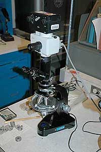
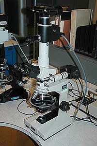
Thin sections are very thin slices of rock used by geologists to help identify minerals. These thin rock specimens are made by slicing a piece of rock using a diamond saw, glueing the specimen to a microscope slide, and then grinding and polishing the specimen to a thickness of around 0.03 mm. The thin sections seen here were prepared by John Schaffer, Thornton, Colorado. Images before 2006 were shot using a Zeiss trinocular polarizing microscope with a Leitz/Wild 35-mm film camera system (above left). More recent thin sections were photographed though a Nikon trinocular polarizing microscope (above right) with a Nikon D-70 digital SLR fitted with a ScopeTronix MaxView Plus eyepiece adapter containing a Plossl lens (photo below).
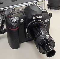 |
Nikon D-70 digital SLR with ScopeTronix MaxView |
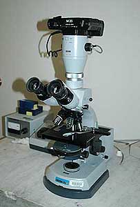
Chemical crystals were prepared using melt crystal formation and precipitation from saturated solutions. Melt crystals were created by placing reagent chemicals (phenol, benzoic acid, citric acid, ascorbic acid, and 4-(2-Pyridylazo)resorcinol-Na monohydrate) on a microscope slide, and heating the slide on a hot plate until the compound melted. A cover slip was then placed on the melted material which crystallized as it cooled. Precipitation crystals were prepared by making a saturated solution of several different inorganic and organic compounds, such as barium chloride, sodium citrate buffer, and potassium acid phthalate. The precipitation crystals were formed by allowing the saturated solutions to evaporate under a cover slip. As the water evaporated, crystals precipitated out of solution. Chemical crystals were photographed using both the Leitz DiaLux-Pol polarizing microscope with the Leitz/Wild film camera, and the Nikon Labophot-Pol polarizing scope with the Nikon D-70 with the ScopeTronix eyepiece adapter.
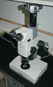
Butterfly and moth wings from the collection of biologist S. Mark Nelson were photographed using an Olympus SZH-10 dissection microscope with a t-tube adaped Nikon FG 35-mm film camera, and focused halogen specimen surface incident lighting. Prepared biological specimen slides from Accu-Scope were photographed through a Zeiss Standard 16 Phase Contrast Microscope with an integrated Zeiss M-35 35-mm film camera. Some of the prepared biological slides were also shot using the Nikon polarizing scope with the Nikon D-70 with the ScopeTronix eyepiece adapter.
|
|
Special thanks to Doug Hurcomb for assistance with the polarizing scopes, and to Denise Hosler for her assistance with the Zeiss phase contrast scope.